
Re-balancing the Tibialis Posterior.
Muscle Management for the Tibialis Posterior
with
Taum Sayers.
This online class presents techniques, protocols, and foundational information relevant to returning the Tibialis Posterior muscle to a state of pain-free function.
The introduction serves to offer an overview of this unique form of therapeutic massage.
The technique videos guide you while you watch and practice the techniques on yourself or with a learning partner. The associated follow-along serves as a condensed refresher.
The class includes 4 sections:
Section 1:
The Corrective Technique for the Tibialis Posterior.
The clay video serves as a visual aid to facilitate your 'tactile' vision.
Section 2: The regional 'Background techniques.'
The video information in section 2 includes fundamental overviews and techniques for reducing regional tensions that often influence the state of the Tibialis Posterior.
Section 3: 'About Muscles.'
This section provides support for a deeper understanding of muscles.
The 'Take your Tools to Work' video information provides examples of positioning when applying techniques to clients.
Section 4: 'The Assignments.'
This section includes videos and relevant information to enhance a foundational understanding of muscles and their essential role in overall health, balance, and pain-free movement.
Once you understand the information presented, click on the exam near the bottom of the class page. When you pass with a 70% or higher score, you receive your Certificate for your 2 CEs.
Note the contact and support links at the bottom of class.
I hope this information serves you and your clients.

Intention and Imagination.
"Repositioning"
Corrective Massage recognizes that muscles, tendons, ligaments, and viscera have optimum anatomical positions, functions, relationships, and movement patterns.
Correcting soft tissue misplacement and distortion is at the core of this amazing work.
Sometimes it's as simple as putting things back where they belong.
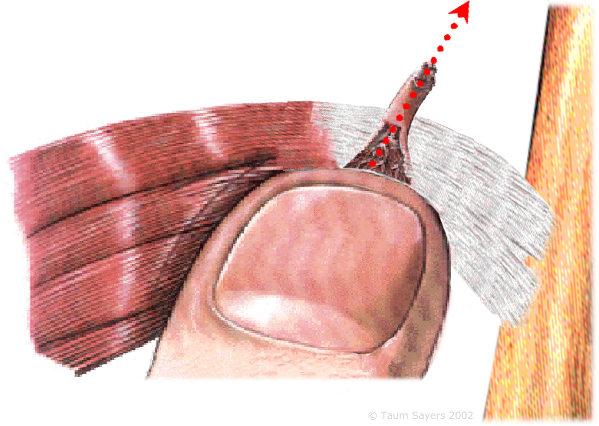 A useful image is one of pushing or nudging dried strings of glue across glass with your fingertips. Consider that the attachment points of the tendon represent a concentration of force in one small area (the entire force of the muscle focuses tension on the bone through the tendon at its attachment). Because of this, the tension/force (pounds per square inch) at the attachment point is higher than at any point in the body of the muscle itself. There are pain receptors at the bone surface (periosteum), which in turn can make the insertion point very sensitive. It is thus helpful to expect these points to be tender and adjust your pressure accordingly. Consider that you are not so much repositioning the fibers to an exact position as much as you are introducing movement towards a balanced situation, thus reducing adhesions and removing distortion. The muscle tissue, upon having the 'duct tape' removed, will most often be able to nestle back to its optimal location. Note that one indication of a misplaced and distorted muscle is when the fibers tactilely 'stand out' with that all too familiar stringy/ropey feeling. Additionally, when muscle fibers are in their most functional position, they usually blend back in with the surrounding fibers and 'tactily disappear'.
The attachment points can serve as an important reference, so after addressing the surrounding regional tensions and distortions within the belly of the tender muscle, you can often return to a previously tender attachment to find it has relaxed. This can also serve to instill confidence in your client that there are benefits to the process.
By applying these techniques, you will have tapped some of the innate intelligence of the body, intentionally encouraging the muscle fiber to release chronic tension by overriding its protective state of contraction via the GTO, thus "reprogramming" it via the nervous system to relax. You will also have encouraged the muscle out of a distorted position and back towards a situation where it has a better opportunity to relax and repair.
This is an excerpt from Taum's article published in the July 2002 issue of Massage Magazine on Mid-Back Tension.*
Click here for the full article: "Intention and Imagination"
In retrospect, a more accurate title might have been
"Defending the Rhomboids."


Tibialis Posterior
ORIGIN: Lateral part of the posterior surface of the tibia, proximal 2/3 of the medial surface of fibula, interosseous membrane, intermuscular septa, and deep fascia.
INSERTION: Tuberosity of navicular bone, plantar surfaces of all three cuneiforms and cuboid, and to bases of the 2nd, 3rd, and 4th metatarsal bones.
ACTION: Inverts and plantar flexes the foot at the ankle. Medial ankle stabilizer.


Terminology
Origin: The beginning site, the more fixed attachment of the muscle. Can be considered as the anchoring point.
Insertion: The movable attachment of the distal end of the muscle.
Entheses: Locations of stress concentration where tendons and ligaments attach to bone.
Superior: Toward the upper part of the structure.
Inferior: Toward the lower part of the structure.
Anterior: Nearer to the front of the body.
Posterior: Nearer to or at the back of the body.
Medial: Near the midline of the body.
Lateral: Farther from the midline of the body.
Proximal: Near the point of attachment, center of the body.
Distal: Away from the trunk, from the center.
External: Towards the surface of the body.
Internal: Away from the surface of and within the body.
Plantar: Refers to the bottom of the foot.
Dorsal: Refers to the top of the foot.
Unilaterally: Contracting on one side.
Bilaterally: Contracting on both sides.
Inversion: To turn inward.
Eversion: To turn outward.
Synergist: Cooperating and supporting.
Antagonist: That which counteracts, opposes.
Agonist: The muscle directly engaged in contraction.
Periosteum: The periosteum is a dense highly innervated membrane composed of fibrous connective tissue that closely wraps (invests) all bone, except that of the articulating surfaces in joints which are covered by a synovial membrane.
The periosteum is the most densely innervated, followed by bone marrow and cortex. It is important to recognize and respect the sensitivity of this membrane. It may well be the most sensitive component in the body.
For more information: Sensory Innervation of Human Bone
"Peri-" is a prefix borrowed from the Greek meaning "around or about" and "-osteum" also comes from the Greek, from "osteon" meaning "bone", so periosteum literally means "around the bone."
Golgi Tendon Organ (GTO): A spindle-shaped structure near the muscular/tendinous junction. This structure is thought to function as a feedback system that monitors muscle tension, inhibits muscle contraction of the agonist, and facilitates contraction of the antagonistic muscle. The purpose of this mechanism is to prevent overuse and damage to the muscle and involved joint.


Additional resources for anatomical clarity:


Re-balancing the Tibialis Posterior.
Muscle Management for the Tibialis Posterior
with
Taum Sayers.
This online class presents techniques, protocols, and foundational information relevant to returning the Tibialis Posterior muscle to a state of pain-free function.
The introduction serves to offer an overview of this unique form of therapeutic massage.
The technique videos guide you while you watch and practice the techniques on yourself or with a learning partner. The associated follow-along serves as a condensed refresher.
The class includes 4 sections:
Section 1:
The Corrective Technique for the Tibialis Posterior.
The clay video serves as a visual aid to facilitate your 'tactile' vision.
Section 2: The regional 'Background techniques.'
The video information in section 2 includes fundamental overviews and techniques for reducing regional tensions that often influence the state of the Tibialis Posterior.
Section 3: 'About Muscles.'
This section provides support for a deeper understanding of muscles.
The 'Take your Tools to Work' video information provides examples of positioning when applying techniques to clients.
Section 4: 'The Assignments.'
This section includes videos and relevant information to enhance a foundational understanding of muscles and their essential role in overall health, balance, and pain-free movement.
Once you understand the information presented, click on the exam near the bottom of the class page. When you pass with a 70% or higher score, you receive your Certificate for your 2 CEs.
Note the contact and support links at the bottom of class.
I hope this information serves you and your clients.
Intention and Imagination.
"Repositioning"
Corrective Massage recognizes that muscles, tendons, ligaments, and viscera have optimum anatomical positions, functions, relationships, and movement patterns.
Correcting soft tissue misplacement and distortion is at the core of this amazing work.
Sometimes it's as simple as putting things back where they belong.
A useful image is one of pushing or nudging dried strings of glue across glass with your fingertips. Consider that the attachment points of the tendon represent a concentration of force in one small area (the entire force of the muscle focuses tension on the bone through the tendon at its attachment). Because of this, the tension/force (pounds per square inch) at the attachment point is higher than at any point in the body of the muscle itself. There are pain receptors at the bone surface (periosteum), which in turn can make the insertion point very sensitive. It is thus helpful to expect these points to be tender and adjust your pressure accordingly. Consider that you are not so much repositioning the fibers to an exact position as much as you are introducing movement towards a balanced situation, thus reducing adhesions and removing distortion. The muscle tissue, upon having the 'duct tape' removed, will most often be able to nestle back to its optimal location. Note that one indication of a misplaced and distorted muscle is when the fibers tactilely 'stand out' with that all too familiar stringy/ropey feeling. Additionally, when muscle fibers are in their most functional position, they usually blend back in with the surrounding fibers and 'tactily disappear'.
The attachment points can serve as an important reference, so after addressing the surrounding regional tensions and distortions within the belly of the tender muscle, you can often return to a previously tender attachment to find it has relaxed. This can also serve to instill confidence in your client that there are benefits to the process.
By applying these techniques, you will have tapped some of the innate intelligence of the body, intentionally encouraging the muscle fiber to release chronic tension by overriding its protective state of contraction via the GTO, thus "reprogramming" it via the nervous system to relax. You will also have encouraged the muscle out of a distorted position and back towards a situation where it has a better opportunity to relax and repair.
This is an excerpt from Taum's article published in the July 2002 issue of Massage Magazine on Mid-Back Tension.*
Click here for the full article: "Intention and Imagination"
In retrospect, a more accurate title might have been
"Defending the Rhomboids."
Tibialis Posterior
ORIGIN: Lateral part of the posterior surface of the tibia, proximal 2/3 of the medial surface of fibula, interosseous membrane, intermuscular septa, and deep fascia.
INSERTION: Tuberosity of navicular bone, plantar surfaces of all three cuneiforms and cuboid, and to bases of the 2nd, 3rd, and 4th metatarsal bones.
ACTION: Inverts and plantar flexes the foot at the ankle. Medial ankle stabilizer.
Terminology
Origin: The beginning site, the more fixed attachment of the muscle. Can be considered as the anchoring point.
Insertion: The movable attachment of the distal end of the muscle.
Entheses: Locations of stress concentration where tendons and ligaments attach to bone.
Superior: Toward the upper part of the structure.
Inferior: Toward the lower part of the structure.
Anterior: Nearer to the front of the body.
Posterior: Nearer to or at the back of the body.
Medial: Near the midline of the body.
Lateral: Farther from the midline of the body.
Proximal: Near the point of attachment, center of the body.
Distal: Away from the trunk, from the center.
External: Towards the surface of the body.
Internal: Away from the surface of and within the body.
Plantar: Refers to the bottom of the foot.
Dorsal: Refers to the top of the foot.
Unilaterally: Contracting on one side.
Bilaterally: Contracting on both sides.
Inversion: To turn inward.
Eversion: To turn outward.
Synergist: Cooperating and supporting.
Antagonist: That which counteracts, opposes.
Agonist: The muscle directly engaged in contraction.
Periosteum: The periosteum is a dense highly innervated membrane composed of fibrous connective tissue that closely wraps (invests) all bone, except that of the articulating surfaces in joints which are covered by a synovial membrane.
The periosteum is the most densely innervated, followed by bone marrow and cortex. It is important to recognize and respect the sensitivity of this membrane. It may well be the most sensitive component in the body.
For more information: Sensory Innervation of Human Bone
"Peri-" is a prefix borrowed from the Greek meaning "around or about" and "-osteum" also comes from the Greek, from "osteon" meaning "bone", so periosteum literally means "around the bone."
Golgi Tendon Organ (GTO): A spindle-shaped structure near the muscular/tendinous junction. This structure is thought to function as a feedback system that monitors muscle tension, inhibits muscle contraction of the agonist, and facilitates contraction of the antagonistic muscle. The purpose of this mechanism is to prevent overuse and damage to the muscle and involved joint.
Additional resources for anatomical clarity:
About Muscles.


Assignments


Taum's approach to manual therapy is primarily influenced by the work of Lauren Berry Sr, Registered Physical Therapist and Structural Engineer.
Structural engineering is a sub-discipline of civil engineering. Structural engineers design the 'bones and muscles' that create the form, shape, and function of structures.
One of Lauren's many unique gifts to the manual therapy world was the concept of soft-tissue balance and distortion.
This is so appropriate:-)
Click here for more information on Lauren.


Terminology
Origin: The beginning site, the more fixed attachment of the muscle. Can be considered as the anchoring point.
Insertion: The movable attachment of the distal end of the muscle.
Entheses: Locations of stress concentration where tendons and ligaments attach to bone.
Superior: Toward the upper part of the structure.
Inferior: Toward the lower part of the structure.
Anterior: Nearer to the front of the body.
Posterior: Nearer to or at the back of the body.
Medial: Near the midline of the body.
Lateral: Farther from the midline of the body.
Proximal: Near the point of attachment, center of the body.
Distal: Away from the trunk, from the center.
External: Towards the surface of the body.
Internal: Away from the surface of and within the body.
Plantar: Concerning the sole of the foot.
Dorsal: Concerning the back of the foot.
Unilaterally: Contracting on one side.
Bilaterally: Contracting on both sides.
Inversion: To turn inward.
Eversion: To turn outward.
Synergist: Cooperating and supporting.
Antagonist: That which counteracts, opposes.
Agonist: The muscle directly engaged in contraction.
Periosteum: The periosteum is a dense highly innervated membrane composed of fibrous connective tissue that closely wraps (invests) all bone, except that of the articulating surfaces in joints which are covered by a synovial membrane.
The periosteum is the most densely innervated, followed by bone marrow and cortex. It is important to recognize and respect the sensitivity of this membrane. It may well be the most sensitive component in the body.
For more information: Sensory Innervation of Human Bone
"Peri-" is a prefix borrowed from the Greek meaning "around or about" and "-osteum" also comes from the Greek, from "osteon" meaning "bone", so periosteum literally means "around the bone."
Golgi Tendon Organ (GTO): A spindle-shaped structure near the muscular/tendinous junction. This structure is thought to function as a feedback system that monitors muscle tension, inhibits muscle contraction of the agonist, and facilitates contraction of the antagonistic muscle. The purpose of this mechanism is to prevent overuse and damage to the muscle and involved joint.

Note that the clearer your question, the clearer my reponse.
Click here to email your question.
or
Call and leave a message: 530-587-6816