
 1055 W Moana Ln #202
Muscle Management® for Rebalancing the Lower Back and Hips.
Saturday, May 23rd, 10-5
Reno
One task to complete your registration:
1 - Click here to email Taum the name you want to be used on your class CE certificate.
This preparation page helps you clarify your knowledge and helps us all focus on the tactile components presented in class.
This is not about memorizing.
Simply watch and read...

Posture and Balance.
Components of Balance/Postural Control
The three sensory systems that are mainly involved in balance or postural control are the somatosensory (proprioception), the visual, and the vestibular systems. In any given situation, the CNS is responsible for prioritizing the information from one system to another in order to maintain balance in different conditions.
The ability to choose and relay the appropriate sensory input for each condition is sometimes referred to as sensory re-weighting. For example, when a person is standing on an unstable surface, the CNS increases sensory weighting to vestibular and visual information and decreases the dependence on surface somatosensory inputs. Likewise, when a person is in the dark, balance control depends primarily on somatosensory and vestibular feedback (de Oliveira, de Medeiros, Frota, Greteers, Conforto, 2008).
The visual system is important for determining orientation and alignment relative to objects and features of the environment. When light strikes the sensory receptors in the retina, it sends impulses to the brain that provide the visual cues necessary to oriented oneself to the surrounding environment.
The somatosensory system combines the sensory inputs of touch and proprioception to provide a sense of body schema and an awareness of the relationship between one’s body and the outside world. Information from the sensory receptors found in skin, muscles and joints, such as the mechanoreceptors (e.g. Pacinian and Meissner corpuscles, Merkle disks, Ruffini endings) and the proprioceptors (e.g. muscle spindle and Golgi tendon organ) help detect the position of body parts in space, and information about joint movement and position.
The vestibular system is part of the inner ear and is responsible for monitoring head position. It also contributes to a sense of equilibrium, which combines balance and orientation in space. Information received from eye movement, as well as head movement, are coordinated through the vestibular system. The vestibular apparatus in the inner ear, sometimes known as the labyrinth is comprised of the cochlea, semicircular canals, utricle and saccule. They function to detect the position and movement of the head and aid in maintaining equilibrium in different contexts.
https://openpub.libraries.rutgers.edu/developmentacrossthelifespan/chapter/posture-and-balance/
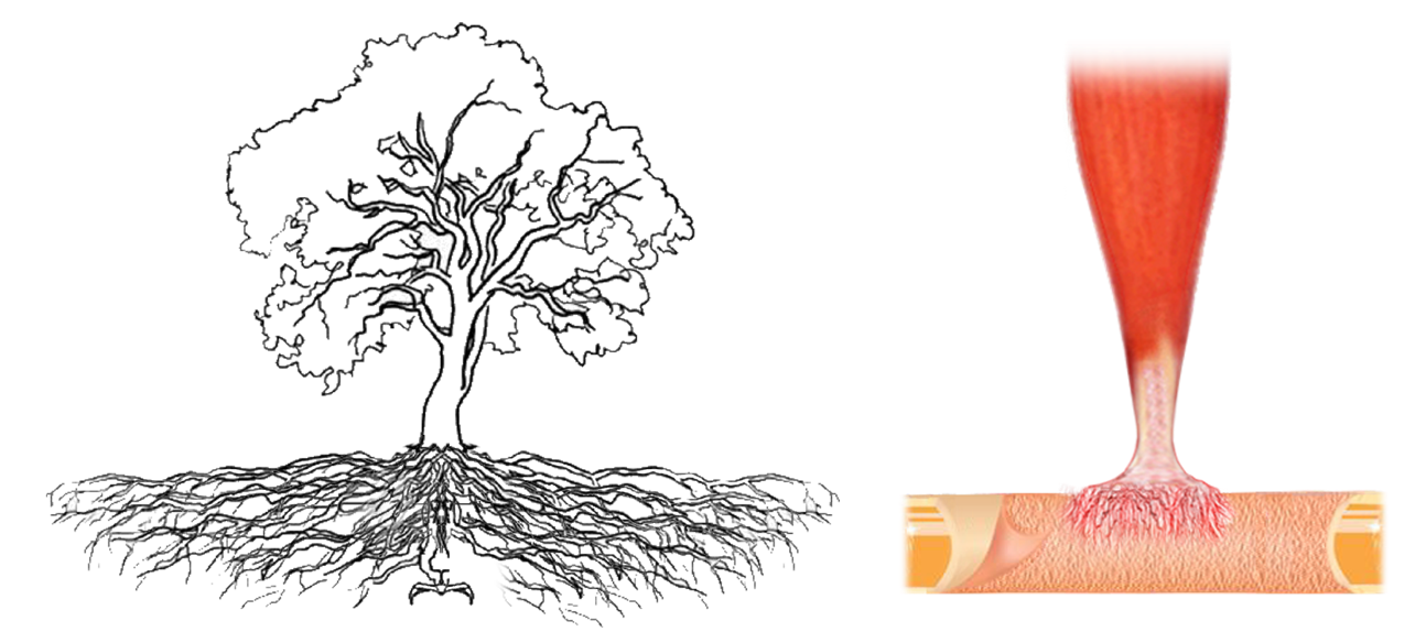
“Like the roots of a tree anchoring it to the earth, the muscles surrounding the lower back and hips create the deep support needed to stand tall and move with ease.”
In this class, we explore the foundational muscles of the lower back and hips—your internal stabilizers—so you can restore balance, alignment, and strength from the inside out.
A helpful image is to imagine the root system as it stabilizes the tree during strong winds.
The body's core muscles we will be focusing on serve a similar purpose.


The quadratus lumborum (QL) is a deep muscle located in the lower back on either side of the lumbar spine. It plays a key role in stabilizing the spine and pelvis and contributes to movement and flexibility in the lumbar region. Often referred to as part of the posterior abdominal wall, the QL is integral to maintaining posture and assisting in lateral flexion of the spine.
Excessive tension in the QL can cause a condition known as 'Quadratus Lumborum Syndrome', resulting in localized lower back pain that may radiate into the pelvis, sacrum, and even the lateral thigh. Overuse, poor posture, prolonged sitting, or heavy lifting can strain the QL, leading to pain and stiffness.
Indications of Quadratus Lumborum Muscle Tension:
- Lower Back Pain (One or Both Sides).
- Hip Pain and Tightness.
- Pain During Breathing.
- Since the QL assists with rib stabilization and can influence breathing, excessive tension in this muscle can make deep breathing uncomfortable or painful. - Pelvic Imbalance or "Hiking" of One Hip.
- A tense QL can pull the hip up on the affected side, causing an uneven pelvis, often called a "hiked" hip. - Referred Pain Patterns.
- Tight QL muscles can create trigger points that refer pain to other areas, often causing discomfort in the hip, buttocks, groin, or outer thigh. This pain can sometimes be mistaken for sciatica, even though it originates from the QL.

http://www.triggerpoints.net/muscle/quadratus-lumborum


 The Ilio-Sacro-Lumbar ligaments (more accurately known as the Iliolumbar ligaments and Sacroiliac ligaments) are critical connective structures in the lower back, helping to stabilize the pelvis, spine, and sacrum. They play significant roles in posture, movement, and load transfer across the pelvis and spine.


Indications of Core musculoskeletal imbalance.
- Relief is short-lived: Pain diminishes by the end of a session, only to resurface soon after.
- Chronic Pain: Persistent discomfort in areas such as the lower back, neck, shoulders, or hips can indicate imbalances or compensations within the musculoskeletal system.
- Postural Misalignment: Visible imbalances such as rounded shoulders, a forward head position, uneven hips, leg length differential, or an exaggerated curvature of the spine (lordosis, kyphosis, or scoliosis).
- Restricted Range of Motion: Difficulty moving joints freely or stiffness in areas like the shoulders, hips, or spine.
- Uneven Muscle Tone: Overdeveloped or underdeveloped muscles on one side of the body, often due to compensatory patterns or poor biomechanics.
- Frequent Injuries: Recurring injuries such as sprains, strains, or overuse injuries, which can result from improper load distribution via ‘Core’ imbalance.
- Fatigue or Weakness: A general sense of muscular fatigue or instability during daily activities or exercise, indicating poor core or foundational strength.
- Balance Issues: Difficulty maintaining balance or coordination during static positions (like standing) or dynamic movements.
- Breathing Patterns: Shallow, chest-dominant breathing instead of diaphragmatic breathing, often linked to tension in the core muscles.
- Gait Abnormalities: Changes in walking patterns, such as limping, uneven stride length, or excessive pronation/supination of the feet.
- Joint Pain or Clicking: Pain, stiffness, or audible clicking in joints like the knees, hips, or shoulders, indicating misalignment or strain.
- Headaches: Tension headaches caused by muscle tightness in the neck, shoulders, or jaw.
- Digestive or Pelvic Dysfunction: Issues such as constipation, bloating, or pelvic floor dysfunction, which can be influenced by improper core muscular engagement and alignment.
Addressing these signs through targeted bodywork can restore balance, improve function, and enhance overall well-being.

Taum's approach to manual therapy is primarily influenced by the work of Lauren Berry Sr, Registered Physical Therapist and Structural Engineer.
Structural engineering is a sub-discipline of civil engineering. Structural engineers design the 'bones and muscles' that create the form, shape, and function of structures.
They also work with and understand the plumbing systems within those structures.
One of Lauren's many unique gifts to the manual therapy world was the concept of soft-tissue balance and distortion.
This is so appropriate:-)
Click here for more information on Lauren.
Terminology
Origin: The beginning site, the more fixed attachment of the muscle. Can be considered as the anchoring point.
Insertion: The movable attachment of the distal end of the muscle.
Entheses: Locations of stress concentration where tendons and ligaments attach to bone.
Superior: Toward the upper part of the structure.
Inferior: Toward the lower part of the structure.
Anterior: Nearer to the front of the body.
Posterior: Nearer to or at the back of the body.
Medial: Near the midline of the body.
Lateral: Farther from the midline of the body.
Proximal: Near the point of attachment, center of the body.
Distal: Away from the trunk, from the center.
External: Towards the surface of the body.
Internal: Away from the surface of and within the body.
Plantar: Refers to the bottom of the foot.
Dorsal: Refers to the top of the foot.
Unilaterally: Contracting on one side.
Bilaterally: Contracting on both sides.
Inversion: To turn inward.
Eversion: To turn outward.
Synergist: Cooperating and supporting.
Antagonist: That which counteracts, opposes.
Agonist: The muscle directly engaged in contraction.
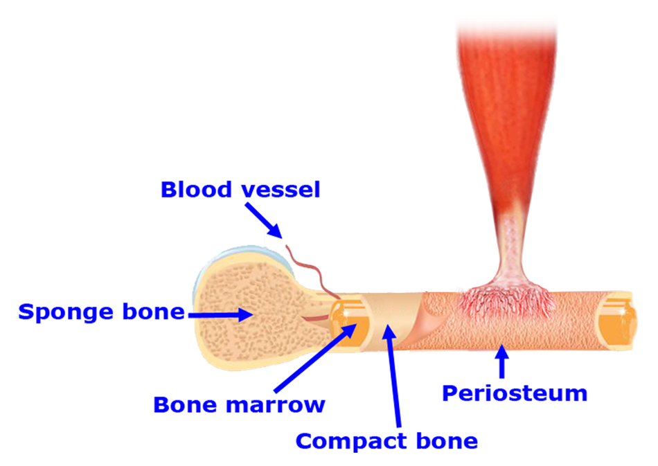
Periosteum: The periosteum is a dense highly innervated membrane composed of fibrous connective tissue that closely wraps (invests) all bone, except that of the articulating surfaces in joints which are covered by a synovial membrane.
The periosteum is the most densely innervated, followed by bone marrow and cortex. It is important to recognize and respect the sensitivity of this membrane. It may well be the most sensitive component in the body.
For more information: Sensory Innervation of Human Bone
"Peri-" is a prefix borrowed from the Greek meaning "around or about" and "-osteum" also comes from the Greek, from "osteon" meaning "bone", so periosteum literally means "around the bone."
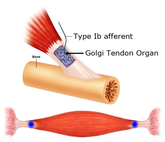
Golgi Tendon Organ (GTO): A spindle-shaped structure near the muscular/tendinous junction. This structure is thought to function as a feedback system that monitors muscle tension, inhibits muscle contraction of the agonist, and facilitates contraction of the antagonistic muscle. The purpose of this mechanism is to prevent overuse and damage to the muscle and involved joint.


Additional resources for anatomical clarity:

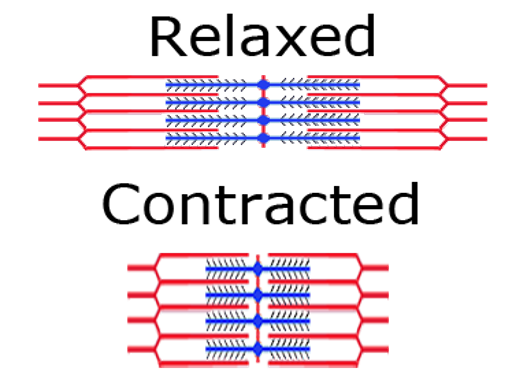
The sarcomere is the fundamental contractile unit of a muscle fiber, and plays a central role in how muscles generate movement.
It is a repeating structural segment within the long, thread-like myofibrils found in muscle cells. Each sarcomere is defined by two Z-lines, which anchor the structure and separate one sarcomere from the next.
Within the sarcomere are thin filaments, primarily made of actin, and thick filaments, made of myosin. During muscle contraction, these filaments slide past one another through a process known as the sliding filament mechanism.
Myosin heads attach to binding sites on actin and pull the filaments inward, shortening the sarcomere. As millions of sarcomeres contract in unison, the entire muscle fiber shortens, resulting in movement.
This sliding interaction between actin and myosin — powered by ATP — is the fundamental basis of voluntary muscle contraction in the body.
Muscle Contraction Process



Terminology
Origin: The beginning site, the more fixed attachment of the muscle. Can be considered as the anchoring point.
Insertion: The movable attachment of the distal end of the muscle.
Entheses: Locations of stress concentration where tendons and ligaments attach to bone.
Superior: Toward the upper part of the structure.
Inferior: Toward the lower part of the structure.
Anterior: Nearer to the front of the body.
Posterior: Nearer to or at the back of the body.
Medial: Near the midline of the body.
Lateral: Farther from the midline of the body.
Proximal: Near the point of attachment, center of the body.
Distal: Away from the trunk, from the center.
External: Towards the surface of the body.
Internal: Away from the surface of and within the body.
Plantar: Refers to the bottom of the foot.
Dorsal: Refers to the top of the foot.
Unilaterally: Contracting on one side.
Bilaterally: Contracting on both sides.
Inversion: To turn inward.
Eversion: To turn outward.
Synergist: Cooperating and supporting.
Antagonist: That which counteracts, opposes.
Agonist: The muscle directly engaged in contraction.
Periosteum: The periosteum is a dense highly innervated membrane composed of fibrous connective tissue that closely wraps (invests) all bone, except that of the articulating surfaces in joints which are covered by a synovial membrane.
The periosteum is the most densely innervated, followed by bone marrow and cortex. It is important to recognize and respect the sensitivity of this membrane. It may well be the most sensitive component in the body.
For more information: Sensory Innervation of Human Bone
"Peri-" is a prefix borrowed from the Greek meaning "around or about" and "-osteum" also comes from the Greek, from "osteon" meaning "bone", so periosteum literally means "around the bone."
Golgi Tendon Organ (GTO): A spindle-shaped structure near the muscular/tendinous junction. This structure is thought to function as a feedback system that monitors muscle tension, inhibits muscle contraction of the agonist, and facilitates contraction of the antagonistic muscle. The purpose of this mechanism is to prevent overuse and damage to the muscle and involved joint.
Additional resources for anatomical clarity:
The sarcomere is the fundamental contractile unit of a muscle fiber, and plays a central role in how muscles generate movement.
It is a repeating structural segment within the long, thread-like myofibrils found in muscle cells. Each sarcomere is defined by two Z-lines, which anchor the structure and separate one sarcomere from the next.
Within the sarcomere are thin filaments, primarily made of actin, and thick filaments, made of myosin. During muscle contraction, these filaments slide past one another through a process known as the sliding filament mechanism.
Myosin heads attach to binding sites on actin and pull the filaments inward, shortening the sarcomere. As millions of sarcomeres contract in unison, the entire muscle fiber shortens, resulting in movement.
This sliding interaction between actin and myosin — powered by ATP — is the fundamental basis of voluntary muscle contraction in the body.
Muscle Contraction Process